|
Research in our laboratory focuses on deciphering the brain circuitry involved in regulating the activity of the gonadotropin releasing hormone (GnRH) neurons, gonadotropin secretion and, ultimately, fertility in all mammals. We are particularly interested in neurons that produce the neuropeptide kisspeptin and project to the GnRH neurons to control their activity. Our goal is to understand how kisspeptin neurons and GnRH neurons integrate a variety of cues (gonadal hormone feedback, internal and external cues) to generate the specific patterns of activity and of gonadotropin secretion that are essential for fertility. We aim to gain insight into how these circuits work under physiological conditions and into how these may become dysregulated under pathological conditions. We use functional approaches such as optogenetics, electrophysiology and calcium imaging, as well as anatomical techniques such as tract-tracing and immunohistochemistry in mice. Research projects are available. Please see our research opportunities page |
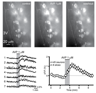
Calcium imaging of kisspeptin neurons in the preoptic area. The top three pictures illustrate GCaMP3 (a genetically-encoded calcium indicator) fluorescence before, during and after the application of arginine vasopressin, a neuropeptide that excites these cells.
The temporal profile of GCaMP3 fluorescence in the cells identified on the images is shown on the bottom left. Population data is shown on the bottom right.
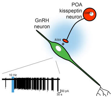
Optogenetic activation of preoptic area (POA) channelrhodopsin-expressing kisspeptin neurons (in red) stimulates GnRH neuron (in green) electrical activity. The trace at the bottom left illustrates a GnRH cell recorded in the cell-attached configuration. A train of blue light pulses delivered at 10 Hz dramatically increases action potential firing (downward deflections).
